Most common fungal skin infections are caused by organisms that feed on keratin and live in the outermost, non-living layer of the skin. They do not invade deeper tissue in healthy skin, which is why they are usually superficial, persistent and quite treatable, and why they often produce a characteristic spreading edge.
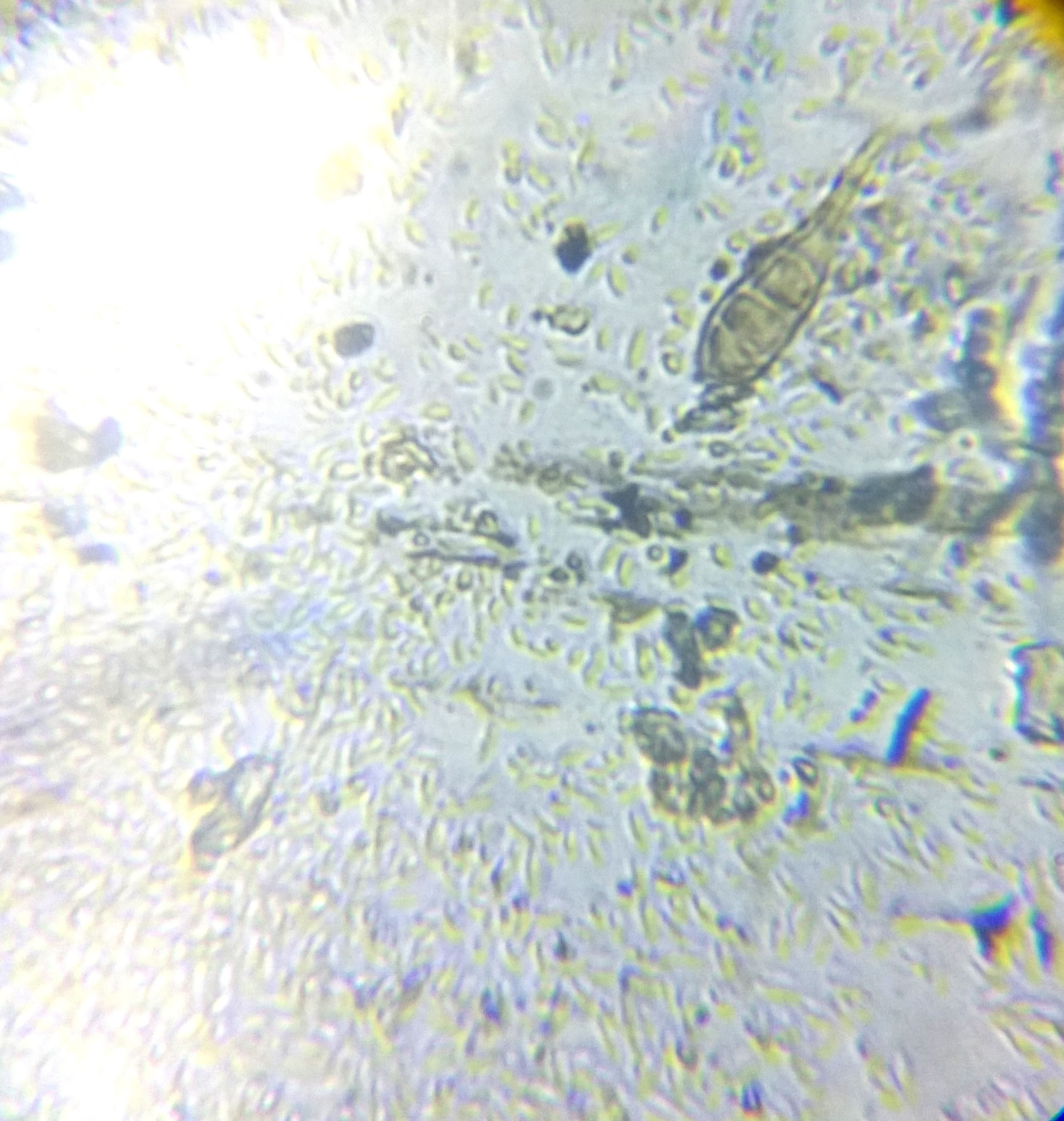

What is actually happening in the skin?
The outermost layer of the skin is not alive. It is a compacted sheet of dead cells packed with keratin, replaced continuously from below, and it forms the barrier that everything else sits under.
A group of fungi called dermatophytes make their living from exactly that material. They digest keratin, which means the outer layer of skin, along with hair and nails, is food. In skin that is otherwise healthy they stay there and do not penetrate the living layers beneath, which is why fungal infections of this kind are described as superficial. The redness and itch are not the fungal organism damaging deeper tissue; they are the immune system responding to its presence at the surface.
Warmth and moisture help fungal growth considerably, which explains the distribution: between the toes, in skin folds, in areas covered by damp clothing. Occlusion matters more than cleanliness.
The spreading pattern follows from how the organism grows. It expands outwards from its starting point, so the leading edge is where the activity is. Behind that edge things are quieter and the skin often looks more normal, producing the ring with a raised rim and a clearer centre. That shape is the origin of the name ringworm, which involves no worm and never has.
Yeasts, including Candida and the Malassezia group, cause a separate set of superficial problems and behave somewhat differently, favouring moist folds and, in the case of Malassezia, oily skin.
How would a dermatologist describe it?
Tinea is the clinical prefix, and it is combined with a Latin word for the site. Tinea corporis means the body, tinea pedis the foot, tinea cruris the groin, tinea capitis the scalp and tinea unguium the nail. Encountering that vocabulary makes an otherwise confusing set of common names suddenly systematic: athlete’s foot and jock itch are locations, not different diseases.
Annular means ring-shaped, and it is used constantly here. The active border may be described as scaly, raised or vesicular, meaning small blisters are present along it. Central clearing describes the quieter middle.
Tinea incognito is the term for a fungal infection whose appearance has been altered by treatment intended for an inflammatory condition, which suppresses the redness without addressing the fungal organism underneath. The name exists because it happens often, and it is a large part of why clinicians prefer to establish what they are looking at before anything is applied.
Scalp involvement is treated as its own category, because it affects the hair shaft as well as the skin and is approached quite differently. That is why a clinician examining a child’s scalp takes it seriously.
What usually happens at a consultation?
The history covers where it started, how it has spread, whether it itches, what has been applied to it, and whether anybody else at home or any animal is affected. Sport, occupation, communal changing facilities and shared footwear all come up. Any previous treatment matters a great deal, because of the altered appearance described above.
The examination looks at the edge of the patch, which is where the diagnostic information is, and generally extends to sites you did not mention. Feet, nails, groin and scalp are checked because a fungal infection travels between sites on the same person, and an untreated reservoir explains many recurrences.
Where the picture is unclear, a clinician may take a sample of skin scrapings, hair or nail clippings for laboratory examination. This is quick, does not hurt, and settles a question that appearance alone sometimes cannot. Results take time to come back, particularly for cultures.
What is worth asking?
Ask whether a sample is worth taking in your case. Establishing what is present, rather than proceeding on appearance, is especially valuable here given how easily a fungal infection is confused with an inflammatory condition.
Ask which sites the clinician checked and what they found. Discovering that your toenails or the skin between your toes are involved changes the picture entirely, and it frequently explains why something on the body keeps returning.
Enquire about the household, including animals. Reinfection from an untreated source at home is one of the commonest reasons these conditions persist, and it is a practical matter a clinician can advise on directly.
Where a laboratory sample is taken, find out when the result is expected and who tells you. These tests can take longer than people assume, and knowing the timescale prevents a lot of unnecessary chasing.
What this page does not do
It does not tell you whether the patch on your skin is fungal. Whether something is fungal is precisely the judgement that appearance makes unreliable and a sample makes solid. No antifungal, product, duration or application schedule is recommended anywhere on this page.
Common questions
Does ringworm involve a worm?
No. The name is purely descriptive of the ring-shaped patch it often produces, and it long predates any understanding of what causes it. Ringworm is a fungal infection, and the misleading name causes enough confusion that clinicians frequently correct it unprompted.
Why does the patch form a ring?
The organism spreads outwards from where it started, so the active, inflamed edge advances while the centre, older and less active, settles. The result is a ring with a raised border and a clearer middle, which is one of the more recognisable patterns in dermatology.
Are athlete's foot and ringworm the same thing?
They are usually caused by the same group of organisms, appearing at different sites. Names such as athlete's foot, jock itch and ringworm describe location rather than distinct diseases, which is why a clinician may examine your feet when you came about somewhere else entirely.
Why is it important not to guess between fungal infection and eczema?
Because some treatments appropriate for inflammatory conditions can alter the appearance of a fungal infection and make it harder to recognise, without resolving it. Clinicians see this often enough to have a name for the altered picture, and it is a good argument for having it looked at.
Can pets pass on fungal skin infections?
Some fungal organisms are carried by animals, and infections acquired from a pet or from farm animals are well recognised. That is why a clinician may ask about animals at home. The question is not idle, and it sometimes explains an otherwise puzzling case.
Sources
- Ringworm and other fungal infections — conditions — NHS (United Kingdom) · accessed
- Patient information on skin conditions — British Association of Dermatologists (United Kingdom) · accessed
Last updated . How this page was written.